Lung cancer remains the leading cause of cancer deaths worldwide, with late stage diagnoses contributing to its high mortality rate. While chest CT scans are the standard for detecting pulmonary nodules, their interpretation is labor intensive and prone to variability. Now, researchers have developed an AI powered vision language model that integrates computer vision with natural language processing to streamline and enhance diagnostic accuracy.
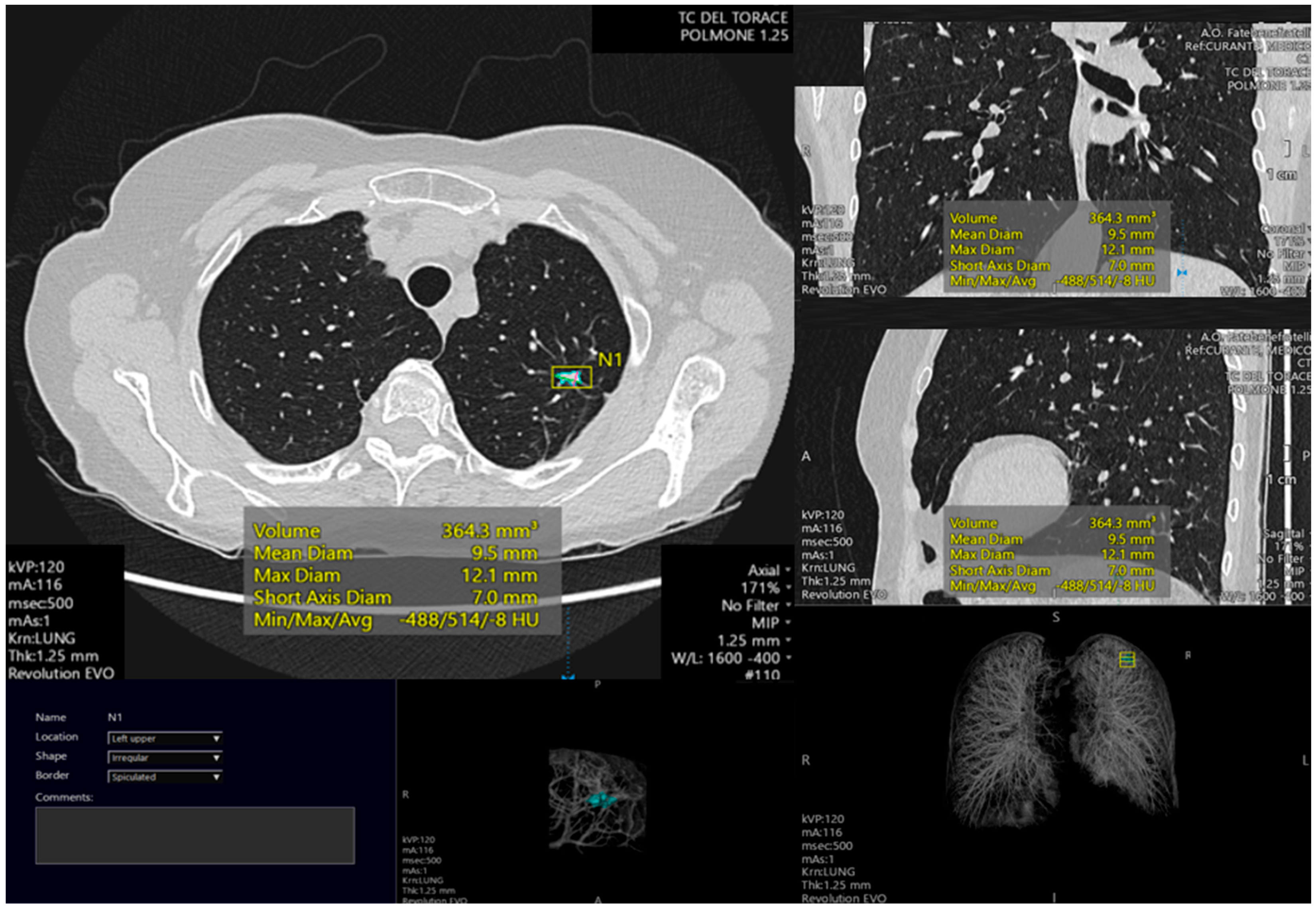
According to a study published in *Nature Medicine*, the model analyzes CT scan images to identify key attributes of pulmonary nodules, such as shape irregularities, margins, and internal patterns, providing radiologists with detailed, contextualized assessments. This technology aims to reduce diagnostic errors and improve early stage lung cancer detection.
What Happened
The research team developed a vision language model that processes CT scan images to generate structured reports for radiologists. The system uses convolutional neural networks to extract visual features from pulmonary nodules and translates these findings into descriptive language, highlighting potential malignancy indicators.
Clinical Significance
Lung cancer’s five year survival rate drops sharply when diagnosed at later stages. Early detection of malignant nodules can lead to timely interventions, such as surgical resection or targeted therapies, significantly improving patient outcomes. The AI model aims to address the global shortage of radiologists and reduce diagnostic variability, particularly in low and middle income countries where access to specialized care is limited.
Deep Dive and Research Findings
The model operates through a multi step process:
- Image Preprocessing: CT scan slices are standardized and segmented to isolate regions of interest, such as pulmonary nodules.
- Feature Extraction: Advanced convolutional neural networks analyze visual characteristics, including size, shape, texture, and density.
- Natural Language Interpretation: Extracted visual data is translated into descriptive language, highlighting critical diagnostic features and potential malignancy indicators.
- Radiologist Integration: The model generates a structured report that radiologists can use to corroborate findings, prioritize cases, and make informed decisions.
Future Outlook and Medical Implications
Researchers are exploring the model’s applicability to other medical imaging modalities, such as mammography and MRI, to broaden its impact. However, challenges remain, including the need for extensive validation across diverse patient populations and integration with existing electronic health record systems. Regulatory bodies, including the U.S. Food and Drug Administration, are increasingly approving AI based diagnostic tools, paving the way for broader clinical adoption.
Patient or Practitioner Guidance
For patients undergoing CT scans for lung cancer screening, the AI model offers a secondary layer of analysis to support radiologists. While the technology enhances diagnostic precision, it is not a replacement for clinical judgment. Radiologists should use AI generated reports as a supplementary tool to prioritize high risk cases and reduce diagnostic variability.
Why This Matters
The global radiology workforce is struggling to meet demand, with many regions facing critical shortages of trained professionals. AI powered tools like this vision language model can help bridge gaps in healthcare delivery, particularly in underserved areas. By improving early detection rates, the technology has the potential to save lives and reduce the burden of lung cancer on healthcare systems.
Key Takeaways
- An AI powered vision language model enhances lung cancer detection in CT scans by analyzing pulmonary nodules with precision.
- The technology reduces diagnostic variability and improves early stage identification, addressing the global shortage of radiologists.
- Researchers are exploring broader applications of the model, including mammography and MRI, to expand its clinical impact.
- Regulatory bodies are increasingly approving AI based diagnostic tools, facilitating their adoption in healthcare settings.
Frequently Asked Questions
How does the AI vision language model improve lung cancer detection?
The model uses convolutional neural networks to analyze CT scan images, extracting visual features from pulmonary nodules and translating these findings into descriptive language. This process highlights potential malignancy indicators and provides radiologists with detailed, contextualized assessments to support their diagnostic decisions.
What are the main challenges in implementing this AI model?
Key challenges include the need for extensive validation across diverse patient populations, integration with existing electronic health record systems, and addressing potential biases in training data. Additionally, ensuring regulatory compliance and ethical deployment remain critical considerations.
Can this AI model replace radiologists?
No, the AI model is designed to augment radiologists' expertise, not replace it. It provides supplementary analysis to reduce diagnostic variability and improve efficiency, but clinical judgment remains essential in patient care.
What types of lung cancer can this AI model detect?
The model is designed to detect pulmonary nodules, which are often the first detectable sign of lung cancer. It can assist in identifying both non small cell lung cancer (NSCLC) and small cell lung cancer (SCLC), though further validation is needed for specific subtypes.
How can patients benefit from this AI technology?
Patients undergoing CT scans for lung cancer screening may benefit from improved early detection rates, particularly in regions with limited access to specialized radiologists. The technology aims to reduce diagnostic errors and prioritize high risk cases for timely interventions.
Medical Review: MedSense Editorial Board


DISCUSSION (0)
POST A COMMENT