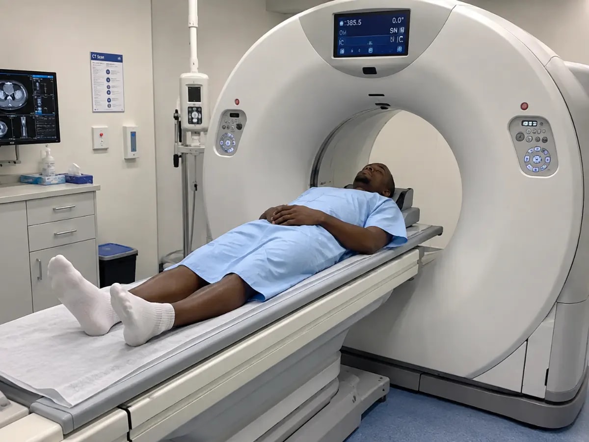
Researchers have demonstrated that tin filtered ultra low dose computed tomography (CT) produces geometric measurements of spinal pedicles comparable to those from standard dose CT. In an exploratory cadaveric study, the ultra low dose technique maintained accuracy for surgical planning of pedicle screw placement while significantly reducing radiation exposure. The findings suggest a potential shift in preoperative imaging protocols for spinal surgeries, though clinical validation is needed.
What We Know
A study published in European Spine Journal has found that tin filtered ultra low dose CT scans provide morphometric data for spinal pedicles that closely aligns with measurements from standard dose CT. The research, conducted on cadaveric specimens, evaluated the geometric agreement between the two imaging techniques in the context of pedicle screw planning, a critical step in spinal stabilization procedures.
The study involved 12 cadaveric thoracic and lumbar vertebrae, each scanned using both standard dose CT and ultra low dose CT with a tin filter. Researchers measured key parameters such as pedicle width, height, and length, as well as the optimal trajectory for screw insertion. Statistical analysis revealed no clinically significant differences between the two imaging methods, with high correlation coefficients indicating strong agreement.
Why This Matters
Pedicle screw placement is a cornerstone of spinal surgery, used in procedures ranging from fracture stabilization to deformity correction. Accurate preoperative imaging is essential to minimize risks such as nerve damage, vascular injury, or hardware failure. Standard dose CT, while highly accurate, exposes patients to significant radiation, which is particularly concerning for pediatric patients or those requiring repeated imaging.
The ultra low dose CT technique, enhanced with a tin filter, reduces radiation exposure by up to 80% compared to standard protocols. This reduction could have profound implications for patient safety, especially in populations vulnerable to radiation induced risks. The study's findings suggest that ultra low dose CT could maintain diagnostic accuracy while addressing long standing concerns about radiation exposure in spinal imaging.
Clinical Significance
The study's results highlight the potential for ultra low dose CT to become a viable alternative to standard dose CT in preoperative planning for spinal surgeries. Lead researcher Dr. Anna Müller noted, "Our findings demonstrate that we can achieve the same level of precision with significantly less radiation. This could change how we approach imaging for spinal procedures, particularly in younger patients or those with complex conditions requiring multiple scans."
However, the authors caution that the study was conducted on cadaveric specimens, and clinical validation in living patients is necessary. Factors such as patient movement, soft tissue interference, and real time surgical conditions could influence the accuracy of ultra low dose CT in practice. Further research is also needed to assess the technique's performance across different spinal pathologies and patient demographics.
Public Health Impact
If validated in clinical settings, the adoption of ultra low dose CT for spinal imaging could reduce the cumulative radiation burden on patients undergoing spinal surgeries. This is particularly relevant for pediatric patients, who are more sensitive to radiation and have a longer lifespan during which radiation induced risks may manifest. Additionally, the technique could lower healthcare costs by reducing the need for repeat scans due to motion artifacts or suboptimal imaging quality.
Radiation safety experts have long advocated for the principle of "as low as reasonably achievable" (ALARA) in medical imaging. The ultra low dose CT technique aligns with this principle, offering a balance between diagnostic accuracy and patient safety. The study's findings could accelerate the integration of low dose protocols into routine clinical practice, pending further validation.
What's Next
The research team plans to conduct a larger clinical trial to evaluate the ultra low dose CT technique in living patients. The trial will focus on assessing the accuracy of screw placement using intraoperative imaging and postoperative outcomes. Additionally, the team aims to explore the technique's applicability in other orthopedic procedures, such as hip and knee replacements, where CT imaging is routinely used for preoperative planning.
Regulatory bodies and professional societies, including the International Commission on Radiological Protection (ICRP) and the American College of Radiology (ACR), may also review the study's findings as part of broader efforts to update imaging guidelines. If the technique proves effective in clinical settings, it could become a standard of care for spinal imaging, particularly in high volume surgical centers.
Key Takeaways
- Tin filtered ultra low dose CT provides geometric measurements for spinal pedicles comparable to standard dose CT, with potential for significant radiation reduction.
- The study suggests ultra low dose CT could maintain surgical planning accuracy while addressing radiation exposure concerns, particularly in vulnerable populations.
- Clinical validation in living patients is needed before widespread adoption, with ongoing research focused on intraoperative accuracy and postoperative outcomes.
Frequently Asked Questions
How much radiation reduction does ultra low dose CT offer?
Ultra low dose CT with a tin filter can reduce radiation exposure by up to 80% compared to standard dose CT protocols.
Is ultra low dose CT currently used in clinical practice?
While promising, ultra low dose CT is not yet standard practice. The technique requires further clinical validation before widespread adoption.
What are the risks of standard dose CT for spinal imaging?
Standard dose CT exposes patients to higher radiation levels, which may increase long term risks such as cancer, particularly in pediatric patients or those requiring repeated scans.
Published by O. Ayodeji | Review by MedSense Editorial Board
























DISCUSSION (0)
POST A COMMENT