Smoking does more than stain teeth and damage lungs. A new study from Ankara University reveals it fundamentally alters the structure and blood supply of palatal tissue, a critical site for dental grafts. Using advanced high frequency ultrasonography, researchers found that heavy smokers develop thicker epithelial layers while suffering reduced vascularization, changes that could complicate oral surgeries and healing processes. The findings, published in Scientific Reports, provide the first detailed imaging evidence of how tobacco use reshapes oral tissue at a microscopic level, offering dentists and periodontists new tools to assess patient risk before procedures.
Clinical Significance
This study breaks new ground by applying high frequency ultrasonography to oral tissue analysis, a technique more commonly used in dermatology and ophthalmology. The ability to visualize epithelial thickness and blood flow in real time gives clinicians an objective way to evaluate graft donor sites before surgery. For heavy smokers, the reduced vascularization observed in the study suggests a higher risk of poor graft integration and delayed healing, critical factors in procedures like gingival grafts or periodontal plastic surgery.
Deep Dive and Research Findings
The research team, led by periodontologist Merve Atak, examined 42 systemically healthy adults, half non smokers and half heavy or chronic smokers, all requiring palatal graft harvesting. Smoking status was confirmed through salivary cotinine testing, a biomarker that provides more accurate exposure data than self reported habits. High frequency ultrasound imaging revealed three key differences in smokers:
- Thicker epithelial layer: The outer tissue layer in the premolar and molar regions measured significantly thicker in smokers, with statistical correlations showing a direct relationship between cigarette consumption and epithelial growth.
- Reduced blood flow: Doppler and B Flow imaging detected lower vascularization in smokers, indicating compromised nutrient delivery to the tissue. This aligns with known effects of nicotine on blood vessel constriction and endothelial function.
- No change in overall thickness: While the epithelial layer thickened, the deeper lamina propria and total tissue thickness remained unchanged, suggesting smoking primarily affects the surface layer rather than the entire tissue structure.
The study also found that age influenced lamina propria and total thickness, but sex showed no significant impact on any measured parameters. These findings challenge the assumption that smoking uniformly thins all oral tissues and instead reveal a more nuanced pattern of localized thickening with underlying vascular damage.
Future Outlook and Medical Implications
The use of high frequency ultrasonography in this study opens new possibilities for preoperative assessment in dental and periodontal procedures. Currently, clinicians rely on visual inspection and patient history to evaluate graft donor sites, but this imaging technique could provide quantitative data to guide treatment planning. For smokers, the reduced vascularization observed may necessitate modified surgical approaches or extended healing protocols to improve graft success rates.
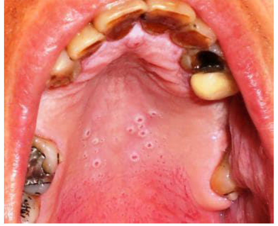
Beyond graft procedures, these findings contribute to the broader understanding of how smoking affects oral health. The epithelial thickening observed could explain why smokers often develop a characteristic leathery texture in their palatal tissue, while the reduced blood flow may contribute to delayed wound healing and increased infection risk after oral surgeries. Future research could explore whether smoking cessation reverses these tissue changes or if the damage persists long term.
Patient or Practitioner Guidance
For dental professionals, this study underscores the importance of thorough preoperative evaluation in smokers. High frequency ultrasonography, though not yet standard in dental practice, could become a valuable tool for assessing tissue quality before graft procedures. Clinicians should consider:
- Incorporating salivary cotinine testing for patients with unclear smoking histories, as self reported data often underestimates true exposure.
- Allowing additional healing time for smokers undergoing palatal graft procedures, given the reduced vascularization observed.
- Discussing these findings with patients to reinforce the oral health risks of smoking beyond tooth discoloration and gum disease.
For patients, particularly those who smoke, these findings highlight another reason to quit. The tissue changes observed in the study suggest that smoking may compromise the success of dental procedures and slow healing after oral surgeries. Patients scheduled for palatal grafts or other periodontal procedures should discuss their smoking status with their dentist, as it may influence treatment planning and recovery expectations.
Key Takeaways
- Heavy smoking thickens the epithelial layer of palatal tissue while reducing blood flow, potentially complicating graft healing and oral surgeries.
- High frequency ultrasonography provides a non invasive way to assess tissue quality before dental procedures, offering objective data beyond visual inspection.
- The study found no difference in overall tissue thickness between smokers and non smokers, challenging assumptions about uniform tissue damage from smoking.
- Reduced vascularization in smokers may necessitate modified surgical approaches or extended healing protocols for optimal graft success.
Frequently Asked Questions
How does smoking affect the palate specifically?
Smoking thickens the outer epithelial layer of palatal tissue and reduces blood flow to the area. This combination may lead to a leathery texture and slower healing after oral procedures, particularly those involving tissue grafts.
What is high frequency ultrasonography, and how is it used in this study?
High frequency ultrasonography is an imaging technique that uses sound waves to create detailed pictures of soft tissues. In this study, it allowed researchers to measure palatal tissue thickness and blood flow without invasive procedures, providing objective data about how smoking alters oral tissue structure.
Should smokers be concerned about dental procedures involving palatal grafts?
The study suggests smokers may face higher risks of poor graft integration and delayed healing due to reduced blood flow in the palate. However, this doesn’t mean smokers can’t undergo these procedures. Dentists may recommend modified approaches, extended healing time, or smoking cessation before treatment to improve outcomes.
Does this study show that smoking cessation can reverse these tissue changes?
The current study did not examine whether quitting smoking reverses the observed tissue changes. However, previous research on smoking cessation shows improvements in blood flow and tissue healing over time. Future studies may explore whether the palatal tissue changes identified here are reversible with smoking cessation.
Medical Review: MedSense Editorial Board


DISCUSSION (0)
POST A COMMENT