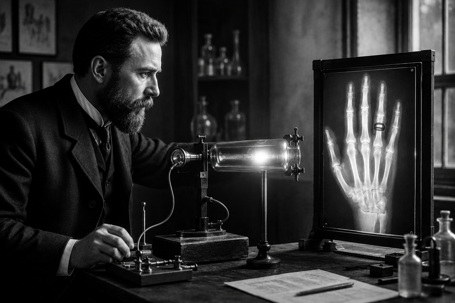
In the quiet hours of a November evening in 1895, a German physicist named Wilhelm Conrad Röntgen made a discovery that would not only redefine science but also transform the practice of medicine for generations to come. While experimenting with cathode ray tubes in his laboratory at the University of Würzburg, Röntgen noticed something extraordinary: a faint glow emanating from a fluorescent screen placed several feet away, even though the tube was encased in thick cardboard. This invisible radiation, which he called "X rays", the "X" signifying the unknown, had the remarkable ability to pass through solid objects, revealing hidden structures within. What began as a curious observation would soon lead to the first Nobel Prize in Physics and lay the foundation for modern diagnostic imaging.
What Are X Rays?
X rays are a form of electromagnetic radiation, similar to visible light but with much shorter wavelengths and higher energy. This higher energy allows X rays to penetrate materials that absorb or reflect visible light, such as human tissue, wood, and metal. When X rays pass through the body, they are absorbed differently by various tissues: bones, which are dense, absorb more X rays and appear white on an image, while softer tissues like muscles and organs allow more X rays to pass through, appearing in varying shades of gray. This differential absorption creates the contrast needed to produce images of internal structures, making X rays an invaluable tool for identifying fractures, infections, tumors, and other abnormalities.
Röntgen’s discovery was groundbreaking not only because it revealed a new type of radiation but also because it demonstrated that invisible forces could be harnessed to see inside the human body without surgery. This capability was unheard of at the time and opened doors to entirely new fields of medical and scientific inquiry.
How the Discovery Unfolded
The story of X rays begins with the study of cathode rays, which were already a subject of fascination in the late 19th century. Cathode rays are streams of electrons produced in a vacuum tube when an electric current is applied. Scientists like Heinrich Hertz and Philipp Lenard had been experimenting with these rays and their properties, including their ability to cause fluorescence in certain materials. Röntgen, a meticulous and curious researcher, was particularly interested in understanding the full range of behaviors exhibited by cathode rays.
On November 8, 1895, Röntgen was working late in his laboratory, as was his habit. He had covered his cathode ray tube with black cardboard to block the visible light and prevent interference with his experiments. Nearby, he placed a fluorescent screen coated with barium platinocyanide, a compound known to glow when exposed to certain types of radiation. As he activated the tube, he observed a faint greenish glow on the screen, even though it was several feet away and shielded from direct light. Intrigued, he moved the screen farther away and noticed that the glow persisted, suggesting that whatever was causing it was not light but something entirely different. Röntgen quickly realized he had stumbled upon a new form of radiation, one that could penetrate solid objects.
Over the next several weeks, Röntgen conducted a series of experiments to understand the properties of this mysterious radiation. He discovered that X rays could pass through paper, wood, and even thin sheets of metal, but were absorbed by denser materials like bones and metals. He also found that X rays could expose photographic plates, creating images of the internal structures of objects and, most astonishingly, the bones of his own hand. On December 22, 1895, Röntgen took the first ever X ray image of his wife Anna’s hand, which clearly showed the bones and her wedding ring. This image, now known as "Hand mit Ringen," became one of the most iconic photographs in the history of science.
Why This Discovery Mattered
The implications of Röntgen’s discovery were immediate and far reaching. Within weeks of his announcement in December 1895, scientists and physicians around the world began experimenting with X rays, recognizing their potential to revolutionize medicine. Before X rays, diagnosing internal injuries or diseases often required invasive procedures or guesswork. Fractures, for example, could only be confirmed through physical examination or exploratory surgery, which carried significant risks. X rays provided a non invasive way to visualize bones and, later, soft tissues, drastically improving diagnostic accuracy and patient outcomes.
Beyond medicine, X rays had profound implications for physics and chemistry. They offered new insights into the structure of atoms and molecules, paving the way for advancements in crystallography and quantum mechanics. Röntgen’s work also inspired further research into other forms of electromagnetic radiation, including gamma rays and ultraviolet light, expanding the boundaries of scientific knowledge.
In 1901, Röntgen was awarded the first Nobel Prize in Physics for his discovery, a testament to the transformative impact of his work. The Nobel Committee recognized not only the scientific significance of X rays but also their potential to benefit humanity. Today, X rays remain a cornerstone of medical imaging, used in everything from routine dental checkups to complex surgical procedures and cancer treatments.
Early Challenges and Ethical Considerations
While the benefits of X rays were undeniable, the early years of their use were not without challenges. The technology was in its infancy, and the equipment was cumbersome and often unreliable. Early X ray machines required long exposure times, which meant patients had to remain still for extended periods, sometimes leading to blurry or unclear images. Additionally, the dangers of radiation exposure were not yet fully understood. Scientists and physicians of the time were unaware of the risks posed by prolonged or repeated exposure to X rays, and many early users suffered from radiation burns or other health issues as a result.
One of the most famous cases of radiation injury involved Thomas Edison’s assistant, Clarence Dally, who worked extensively with X rays in the late 19th and early 20th centuries. Dally developed severe radiation burns on his hands and arms, which eventually led to his death in 1904. His case highlighted the need for safety protocols and protective measures when working with X rays, a lesson that would shape the future of radiology and medical imaging.
Over time, advancements in technology and a deeper understanding of radiation safety led to the development of more sophisticated and safer X ray equipment. Protective shielding, reduced exposure times, and improved imaging techniques have made X rays a routine and safe diagnostic tool in modern medicine.
Legacy and Modern Applications
Röntgen’s discovery of X rays marked the beginning of a new era in science and medicine, one that continues to evolve today. The field of radiology has grown exponentially, with X rays serving as the foundation for a wide range of imaging techniques. Computed tomography (CT) scans, mammography, and fluoroscopy are all descendants of Röntgen’s original discovery, each offering unique insights into the human body.
X rays are also used in fields beyond medicine. In archaeology, X ray fluorescence is used to analyze artifacts and determine their composition without damaging them. In art conservation, X rays help reveal hidden layers in paintings or detect forgeries. In security, X ray scanners are employed to inspect luggage and cargo for hidden threats. The versatility of X rays has made them an indispensable tool across multiple industries, underscoring the enduring impact of Röntgen’s work.
Beyond their practical applications, X rays have also captured the public imagination, symbolizing the power of scientific discovery to unlock the mysteries of the natural world. Röntgen himself was a humble and private individual who shunned the spotlight, preferring to let his work speak for itself. Despite his reluctance to seek fame, his contributions have left an indelible mark on history, reminding us of the profound impact that curiosity and perseverance can have on the world.
Key Takeaways
- X rays are a form of high energy electromagnetic radiation that can penetrate solid objects, revealing internal structures like bones and organs.
- Wilhelm Conrad Röntgen discovered X rays accidentally in 1895 while experimenting with cathode ray tubes, leading to the first Nobel Prize in Physics in 1901.
- The discovery revolutionized medicine by providing a non invasive way to diagnose fractures, infections, and other internal conditions.
- Early use of X rays posed radiation risks, which were later mitigated through advancements in technology and safety protocols.
- X rays have applications beyond medicine, including archaeology, art conservation, and security, demonstrating their versatility and enduring relevance.
Frequently Asked Questions
How did Röntgen come up with the name 'X rays'?
Röntgen named the newly discovered radiation "X rays" because the "X" symbolized the unknown. At the time of his discovery, the nature of the radiation was not understood, and he chose the term to reflect its mysterious properties.
What was the first X ray image ever taken?
The first X ray image ever taken was of Röntgen’s wife Anna’s hand, captured on December 22, 1895. The image clearly showed the bones of her hand and her wedding ring, demonstrating the ability of X rays to reveal internal structures.
How did the discovery of X rays impact medicine?
The discovery of X rays transformed medicine by providing a non invasive method to visualize internal structures, such as bones and organs. This allowed for more accurate diagnoses of fractures, infections, tumors, and other conditions, reducing the need for exploratory surgery and improving patient outcomes.
What were the early risks associated with X rays?
Early users of X rays were unaware of the risks posed by radiation exposure. Prolonged or repeated exposure could lead to radiation burns, skin damage, and other health issues. Cases like that of Clarence Dally, who died from radiation related injuries, highlighted the need for safety protocols and protective measures.
How are X rays used today beyond medical imaging?
Beyond medical imaging, X rays are used in archaeology to analyze artifacts, in art conservation to study paintings, and in security to inspect luggage and cargo. They are also employed in industrial settings to inspect materials for defects and in scientific research to study the structure of molecules and crystals.
Medical Review: MedSense Editorial Board
DISCUSSION (0)
POST A COMMENT